A PET (Positron Emission Tomography) scan is a test done in the Nuclear Medicine area of the Radiology Department. Most often, doctors will order a PET scan to look for tumors. However, PET scans are also used to diagnose infection or inflammation, and to evaluate your child if he has seizures or other neurological concerns.
For a PET scan, a glucose- or sugar-based radioactive medicine is given to your child through an IV. This IV medicine moves through the body to any organ that utilizes sugar such as the brain, heart, liver, and kidneys. The PET scan provides a picture of these parts of your child’s body and also shows cells that are actively using sugar in areas of infection or a growing tumor.
For a whole body PET scan, your child must follow special instructions before arriving for the scan. She cannot have anything to eat or drink for 4 hours before arriving. She should also not take any medications containing glucose or sugar. If these instructions are not correctly followed, it will decrease the image quality and the scan will be rescheduled.

Once you and your child check in and register in the Radiology Department, a nurse will weigh your child and bring you to a small preparation room. There, the nurse will complete a medical history while your child lies on a bed with warm blankets. When a person is cold, some fat in the body can take in the radioactive medicine that will be given for the scan, affecting the quality of the images. Warming your child prevents this from happening and helps make the images clearer. A medication called Fentanyl may also be given to lessen the amount of the radioactive medicine that is taken in by some fat in the body.
After your child has been warmed and the Fentanyl has been given, the radioactive glucose medicine is put through the IV. Once this medicine is given, it is important that your child lie as still and quiet as possible. Having your child be calm and still helps the radioactive medicine appropriately be taken in by the body. Your child can watch a movie or take a nap during this time. After 45 minutes of quietly resting, your child will be ready to have the PET scan pictures taken.
If your child is getting a PET scan of only the brain, there are no special instructions to follow before the appointment. Only the radioactive glucose medicine is given through a small butterfly injection. Your child will then lie relatively still and quiet for 30 minutes before starting the PET scan.

For a whole body PET scan, the pictures can take 15 to 30 minutes. For a brain PET scan, the pictures take about 10 minutes. Your child will need to lie still in the PET scanner while the pictures are taken. Movie goggles are available for your child to use during the scan and you are able to stay in the room. Younger children may require sedation in order to keep them still for the pictures.
The PET scan camera takes pictures of the radioactive medicine in the body. The same camera can be used to take a quick CT scan, which will be used to help increase the quality of the PET scan images. For the CT part of the scan, you’ll be given a lead apron to wear to shield you from the low dose radiation. After the CT scan, the lead aprons can be taken off, since the camera will not produce any radiation during the PET scan.
Once the nuclear medicine physician checks the images, the scan is complete. It takes about 24 to 48 hours for the ordering doctor to get the final results of the PET scan. The nuclear medicine care team understands that the results of a PET scan are very important to you and your child’s care and will do everything they can to ensure the highest quality images and the most supportive experience possible.
Story contributed by Bessie Ganim, Nuclear Medicine Technologist and Susan Sharp, MD. Edited by Catherine Leopard.
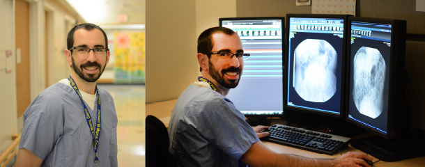
Alex is a radiologist and the Neil D. Johnson Chair of Radiology Informatics. In this role, he helps to manage the information systems used by the Radiology department. Clinically, Alex is the Assistant Director of thoracoabdominal imaging. His research interests include liver disease, liver tumors, inflammatory bowel disease, and appendicitis.